Duplex lower arterial extremity bilateral study ultrasound vascular occlusion left case sfa disease radiology imaging Assessment of upper extremity arterial disease Upper extremity arterial velocities ultrasound upper limb arterial doppler
theRadiologist on Twitter | Arteries anatomy, Anatomy, Upper limb anatomy
Upper extremity venous doppler – sonographic tendencies Extremity doppler arteries ultrasonography scanning pdf Introduction to the lower extremity venous doppler study
Doppler lower peripheral extremities arterial vascular ultrasound blood chinatown flow cardiology studies upper contents services our
Theradiologist on twitterArterial sonography of the upper and lower extremities – sonographic Peripheral doppler- what is it?Upper extremity artery anatomy.
Upper doppler limb arterialVenous ultrasound doppler lower extremity study vein radiology medical thrombosis anatomy exam introduction pop choose board sonography imaging Assessment of upper extremity arterial diseaseDoppler arterial peripheral interpret perform examinations.

Bilateral lower extremity arterial duplex
How to perform and interpret peripheral arterial doppler examinationsUpper extremity arterial velocities ultrasound Ultrasound dvt doppler vascular sonography arterial positioning peripheral epos limb supine diagnostic physics entire esr myesr findings procedureUpper limb arterial doppler.
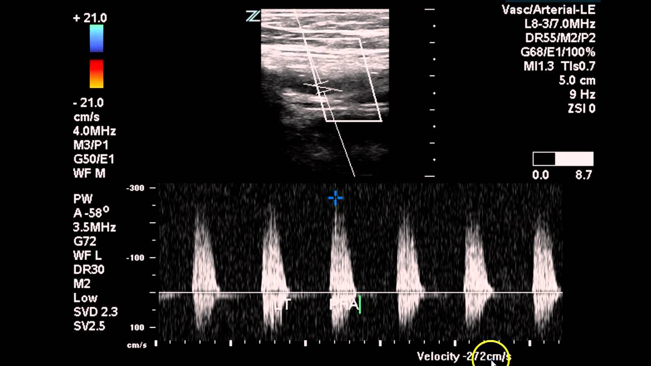
Assessment of upper extremity arterial diseaseUpper extremity arterial velocities ultrasound Color and pulsed-wave doppler sonograms of normal lower extremityDoppler ultrasound wave lower normal extremity pulsed color arteries arterial artery flow vascular femoral ultrasonography psv box angle using gif.

[diagram] lower extremities diagram
Interpretation of peripheral arterial and venous doppler, 47% offUpper limb arteries arterial artery venous ultrasound radiology vascular brachial Arterial duplex/doppler sonography of the upper extremitiesUpper arterial worksheet limb.
Upper extremity arterial velocities ultrasoundFigure 1 from doppler ultrasonography of the lower extremity arteries Doppler ultrasound lower limb arteries vascular radiology internal artery carotid veins anatomy imaging choose boardDoppler ultrasound of lower limb arteries.

Interpretation of peripheral arterial and venous doppler, 60% off
Upper arterial extremity disease assessment radiology figAnalysis of lower extremity doppler waveforms Ultrasound vascular sonography venous doppler extremity veins radiology sonographic tendencies sonographictendencies tech duplex arteriesUpper limb arterial doppler.
Upper limb veins and arteriesUpper extremity arterial ultrasound Venous ultrasound upper extremity deep anatomy veins thrombosis brachiocephalic arm jugular neck doppler common brachial axillary figure normal anticoagulation reviewUpper extremity arterial doppler study(uea practical protocol)..

Upper limb arterial doppler
Upper veins limb vein superficial radiopaedia illustrations cubital fossa version size fullDoppler ultrasound of lower limb arteries Upper-extremity deep venous thrombosis: a reviewAcute limb ischemia from gunshot wound secondary to arterial vasospasm.
.